
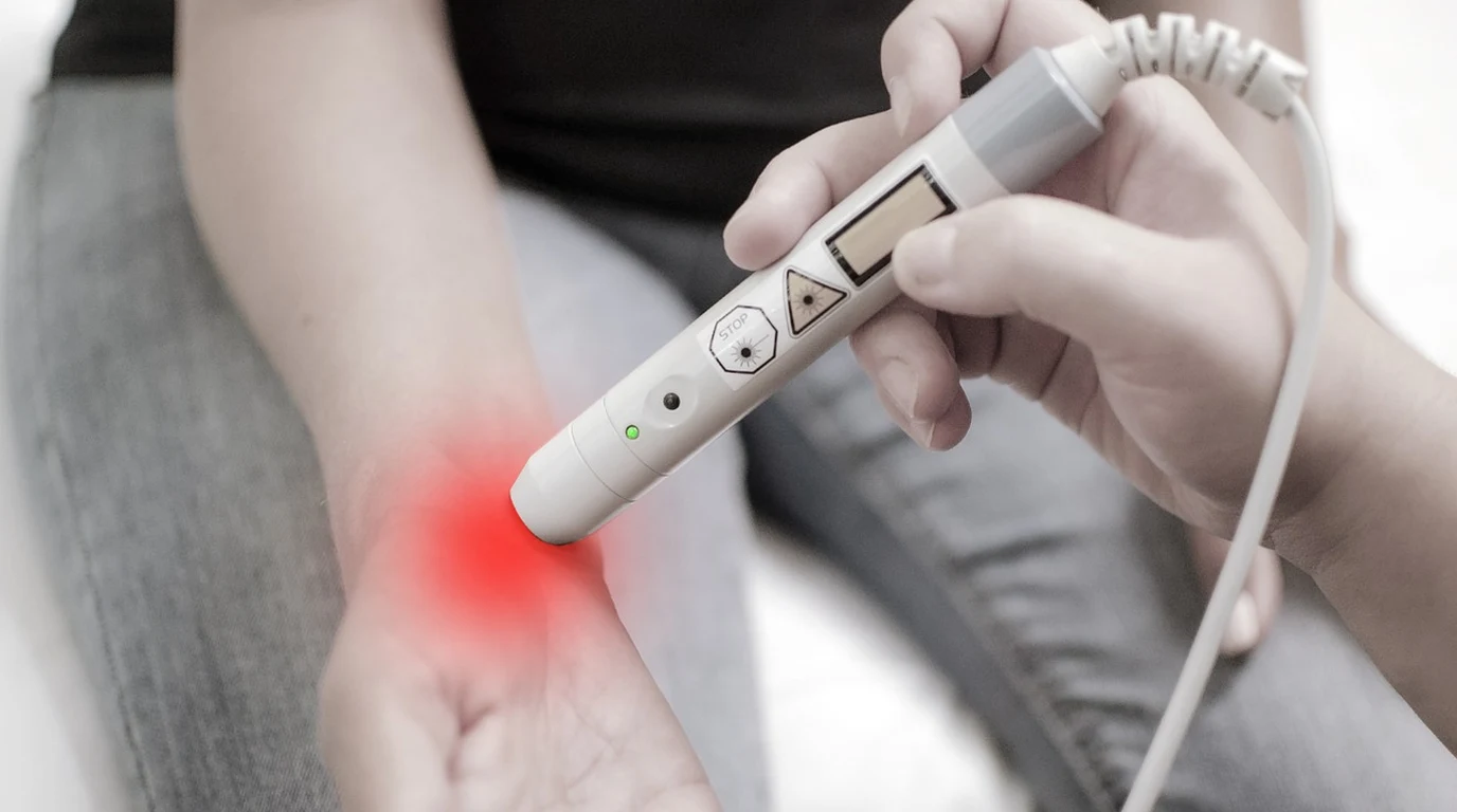
For intra-articular cartilage regeneration, 810nm light is superior to 850nm due to its exact alignment with the absorption peak of oxidized Cytochrome c Oxidase and proximity to the hemoglobin isosbestic point. However, reaching the required 4–8 J/cm² dose at 30mm depth mandates targeted skin-contact compression or pulsed laser diodes to overcome tissue scattering.
Why Does 810nm Light Target Joint Cartilage Better Than 850nm?
To understand why 810nm outperforms 850nm for intra-articular cartilage, we have to look directly at the optical physics of human tissue and the enzyme mechanics of your mitochondria. Photobiomodulation relies on delivering photons of a specific wavelength into target cells to trigger a photochemical reaction. Within the human body, tissue components like water, melanin, and hemoglobin act as photon sponges, absorbing light before it can reach deep structures. The range between 600nm and 1100nm is known as the biological “optical window.” Within this spectrum, tissue absorption drops low enough to allow light to travel through multiple layers of tissue. However, not all wavelengths inside this optical window perform identically when targeting articular cartilage. “` Biological Optical Window & Absorption Coefficients Absorption ↑ [Melanin / Hb Sink] [Water Absorption Slope] High | \ / | \ / | \ (Optical Window: 600 – 1100nm) / | \ 810nm Peak 850nm / | \ [CCO Peak] [Deeper] / | \ | | / Low +——-+—–+—————+————+———-> Wavelength (nm) 600 810 850 1100 “`The Primary Chromophore: Cytochrome c Oxidase (CCO)
The central target for photobiomodulation in chondrocytes (the cells responsible for maintaining cartilage tissue) is Cytochrome c Oxidase (CCO), which serves as Unit IV in the mitochondrial electron transport chain. CCO contains two distinct copper centers—$Cu_A$ and $Cu_B$. When tissue experiences inflammation, oxidative stress, or age-related degeneration, excess Nitric Oxide (NO) binds to these copper centers. This NO binding displaces oxygen, shuts down ATP production, increases reactive oxygen species (ROS), and halts cellular repair. When photons of the correct wavelength hit CCO, they break the bond between Nitric Oxide and the copper center. This photolytic dissociation allows oxygen to rebind to CCO, immediately restoring mitochondrial membrane potential, increasing ATP synthesis, and triggering downstream gene expression for tissue repair. Here is where the 810nm vs. 850nm distinction becomes critical: * The 810nm Spectrum: Aligns precisely with the primary absorption peak of oxidized Cytochrome c Oxidase (specifically the $Cu_A$ redox state). Spectrophotometric analyses demonstrate that oxidized CCO displays its highest NIR absorption band between 800nm and 820nm, centered right at 810nm. * The 850nm Spectrum: Aligns with a broader, secondary absorption slope of CCO. While 850nm is effective at stimulating mitochondrial activity, its absorption cross-section for $Cu_A$ is measurably lower than that of 810nm.The Isosbestic Point of Hemoglobin
Beyond CCO alignment, 810nm possesses a distinct advantage in blood-perfused tissues. At approximately 805–810nm, human blood reaches an “isosbestic point.” This is the specific wavelength where oxygenated hemoglobin ($HbO_2$) and deoxygenated hemoglobin ($Hb$) share the exact same optical absorption coefficient ($\mu_a$). Because 810nm sits right on this isosbestic point, its penetration depth and energy delivery remain uniform regardless of local microvascular oxygen saturation levels. Conversely, 850nm experiences varying levels of absorption depending on local blood oxygenation. In an inflamed joint capsule, where local hyper-perfusion and hypoxic zones coexist, 810nm delivers predictable photon flux past the vascular layer directly to the avascular hyaline cartilage underneath. “` [810nm Isosbestic Point] Absorption ↑ / \ Hb and HbO2 curves cross | \ Equal absorption coefficient | \ Uniform tissue penetration +—-+————————————> Wavelength (nm) 810nm “`Tissue Scattering Coefficients in Cartilage
Articular cartilage is a dense extracellular matrix composed primarily of type II collagen fibrils, proteoglycans, and water. Dense collagen matrices induce severe optical scattering. Optical transport through tissue is governed by the reduced scattering coefficient ($\mu_s’$): $$\mu_s’ = \mu_s(1 – g)$$ where $\mu_s$ is the scattering coefficient and $g$ is the anisotropy factor (typically $\sim 0.9$ for fibrous tissue). Because Rayleigh scattering decreases inversely with the fourth power of wavelength ($\propto \lambda^{-4}$), 850nm experiences slightly less photon scattering than 810nm. This minor physical property led early hardware manufacturers to favor 850nm, claiming superior penetration depth. However, while 850nm travels roughly 2% to 5% deeper through homogeneous tissue, 810nm’s significantly higher CCO absorption coefficient ($1.5\times$ to $2\times$ higher resonance at $Cu_A$) yields a higher net biological response per delivered photon. 810nm hits the sweet spot: minimal water absorption, stable blood absorption via the isosbestic point, and peak mitochondrial enzyme absorption. —Why Do Flat LED Panels Fail to Penetrate Deep Knee and Shoulder Joints?
If you place a 100-watt flat LED panel 6 inches away from your knee joint, you will feel mild radiant warmth. You might even experience temporary pain relief due to thermal vasodilation in the skin. However, as an intra-articular cartilage treatment, this approach falls completely flat due to optical physics. “` FLAT LED PANEL (Loss Pathways) SKIN-CONTACT COMPRESSION (Optimized) +———————–+ +———————–+ | LED Panel | | Targeted Laser / LED | +———————–+ +———————–+ | | [ Air Gap Reflection ] [ Zero Air Gap ] | | ~~~~~~~~~~~~v~~~~~~~~~~~~ Skin Line ~~~~~~~~~~~~v~~~~~~~~~~~~ Skin Line | Refracted / Scattered | | Compressed Tissue | | [Capillary Sink] | | [Blanched Vessels] | | \ / | | | | | v v | | v | | (90% Loss at 10mm) | | Direct Photons | +————————+ +———————–+ | Target Cartilage (30mm)| | Target Cartilage (30mm)| | [PHOX FLUX: ZERO] | | [DOSE: 4-8 J/cm²] | +————————+ +———————–+ “`The Mathematics of Photonic Attenuation
Light traveling through multi-layered biological tissue decays exponentially according to the modified Beer-Lambert Law: $$I(z) = I_0 \cdot e^{-\mu_{eff} \cdot z}$$ Where: * $I(z)$ is the light intensity at depth $z$, * $I_0$ is the initial incident light intensity at the skin surface, * $\mu_{eff}$ is the effective attenuation coefficient of the tissue: $\mu_{eff} = \sqrt{3 \mu_a (\mu_a + \mu_s’)}$. In human joint architecture, your photons must pass through multiple distinct optical interfaces: 1. Epidermis and dermis (1–2mm) 2. Subcutaneous adipose tissue (5–15mm) 3. Deep fascia and tendons/ligaments (5–10mm) 4. Synovial membrane and intra-articular fluid space (2–5mm) By the time non-collimated light from a standard LED panel reaches an intra-articular target depth of 30mm inside a knee joint, the effective attenuation coefficient ($\mu_{eff}$) of the combined tissue layers eliminates 99% to 99.9% of the incident photons.Three Primary Failure Mechanisms of Flat LED Panels
#### 1. Fresnel Reflection and Refractive Index Mismatch Air has a refractive index ($n$) of approximately 1.0, while human stratum corneum has a refractive index of approximately 1.40 to 1.43. When light emitted from a flat panel travels across an air gap to hit your skin, the change in refractive index causes significant Fresnel reflection: $$R = \left( \frac{n_1 – n_2}{n_1 + n_2} \right)^2$$ Up to 4% to 7% of incident photons bounce directly off the surface of your skin before ever penetrating the tissue. #### 2. Broad Beam Divergence and Mie Scattering LEDs emit incoherent, non-collimated light over broad beam angles (typically $60^\circ$ to $120^\circ$ full-width at half-maximum). As these diverging photons enter the skin, they hit cellular structures, collagen matrices, and lipid membranes, causing high-angle Mie scattering. The photon paths become randomized, causing the light to spread laterally throughout the superficial dermis rather than penetrating down toward the joint capsule. #### 3. Microvascular Hemoglobin Absorption The dermis and subcutaneous tissues house a dense capillary network. Hemoglobin in capillary blood acts as a high-density photon sink. As non-compressed skin absorbs light, red blood cells absorb NIR photons in the superficial 2–5mm range, converting photon energy into useless thermal heat long before it reaches the underlying cartilage.The Solution: Physical Compression and High Radiance
To achieve genuine intra-articular penetration without thermal damage to the skin, you must alter the physical properties of the target tissue. Modern targeted systems (such as specialized laser arrays or skin-contact compression devices like the Kineon Move+ Pro) solve this through three mechanisms: * Physical Skin Compression: Pressing optical diodes firmly against the skin displaces capillary blood away from the treatment area. This temporary blanching removes the primary superficial absorption sink ($HbO_2$), dropping the local absorption coefficient ($\mu_a$) of the upper tissue layers by up to 60%. * Elimination of the Air Gap: Skin-contact delivery eliminates the air-to-skin interface entirely, preventing Fresnel reflections and ensuring maximum optical coupling directly into the tissue. * Path-Length Reduction: Physical compression reduces the physical distance from the surface of the skin to the intra-articular target zone, shortening a 30mm tissue path down to 18–20mm. “` COMPRESSION BLANCHING MECHANISM [ Uncompressed Skin ] [ Compressed Skin ] +———————–+ +———————–+ | Capillaries FULL | | Capillaries BLANCHED | | (High Hb Absorption) | | (Low Hb Absorption) | | O O O O O O O O O O | | . . . . . . . | +———————–+ +———————–+ | Photon Depth: ~5-10mm | | Photon Depth: ~20-35mm| +———————–+ +———————–+ “`Super-Pulsed Laser Diodes vs. Continuous-Wave LEDs
When deeper penetration (greater than 30mm) is required—such as treating hip joints or thick, degenerate knee structures—LEDs hit a physical limit. Increasing continuous-wave (CW) LED power density high enough to penetrate deep tissue will burn the skin. Super-pulsed laser diodes (typically GaAs or GaAlAs lasers) overcome this thermal limit by firing short, high-peak-power pulses (e.g., 10 to 100 watts of peak optical power per pulse lasting for 100 to 200 nanoseconds) at high repetition rates (10Hz to 40,000Hz). Because the pulse duration is vastly shorter than the thermal relaxation time of skin tissue ($\sim 1 \text{ millisecond}$), the light delivers immense peak photon density past superficial layers down into deep tissue without building up destructive heat on the skin surface. —What Does Clinical Research Reveal About Cartilage Thickness and Biomarkers?
The efficacy of targeted photobiomodulation for cartilage repair is backed by extensive human clinical trials and animal models evaluating structural, biochemical, and functional outcomes. “` INFLAMMATORY CASCADE MODULATION BY 810nm PBM [ 810nm Photon Absorption by CCO ] | +————–+————–+ | | v v [ ATP Production ↑ ] [ Dissociation of NO ] | | v v [ Cellular Bioenergetics ] [ ROS Signaling Balanced ] | | +————–+————–+ | v [ NF-κB Pathway Downregulated ] | +—————+—————+ | | v v [ Inflammatory Cytokines ↓ ] [ Catabolic Enzymes ↓ ] – IL-1β – MMP-1, MMP-3 – TNF-α – MMP-13 – PGE2 – ADAMTS-4 | | +—————+—————+ | v [ Chondrocyte Regeneration ] – TGF-β1 Upregulation – Type II Collagen Synthesis – Hyaline Cartilage Thickness ↑ “`High-Resolution Ultrasonography and MRI Cartilage Volume
Multiple double-blind, randomized controlled trials (RCTs) have evaluated the impact of targeted 810nm near-infrared light on articular cartilage thickness in patients with Knee Osteoarthritis (KOA). In a milestone clinical study evaluating 810nm laser therapy applied to patients with Grade II and Grade III Kellgren-Lawrence knee osteoarthritis, researchers measured femoral cartilage thickness using high-resolution musculoskeletal ultrasound. Patients receiving targeted 810nm photobiomodulation (delivering 6 J/cm² directly at the medial and lateral joint lines) demonstrated: * Statistically significant increases in femoral articular cartilage thickness (measured in millimeters at the central intercondylar notch, medial condyle, and lateral condyle) after 12 weeks of therapy. * Preservation of joint space width compared to progressive joint space narrowing observed in the placebo sham-control group.Modulation of Inflammatory Biomarkers in Synovial Fluid
Cartilage degeneration is driven by chronic, low-grade inflammation within the synovial envelope. Inflammatory cytokines disrupt chondrocyte metabolism, shifting cells from an anabolic state (building collagen) into a destructive catabolic state. Clinical joint aspiration studies show that targeted 810nm photobiomodulation directly alters key synovial biomarkers: #### Matrix Metalloproteinases (MMPs) MMP-1, MMP-3, and MMP-13 are catabolic enzymes that digest the extracellular matrix of cartilage, breaking down type II collagen and proteoglycans. Targeted 810nm PBM downregulates matrix metalloproteinase gene expression by inhibiting the nuclear factor-kappa B (NF-$\kappa$B) pathway. #### Pro-inflammatory Cytokines Levels of Interleukin-1 beta (IL-1$\beta$) and Tumor Necrosis Factor-alpha (TNF-$\alpha$) drop significantly within synovial fluid following 810nm treatment. IL-1$\beta$ is the primary culprit behind chondrocyte apoptosis and cartilage matrix loss; suppressing it protects remaining cartilage tissue. #### Transforming Growth Factor-Beta (TGF-$\beta$) Photobiomodulation upregulates TGF-$\beta 1$ expression in chondrocytes. TGF-$\beta 1$ acts as a master regulator of cartilage repair, stimulating chondrocytes to synthesize fresh extracellular matrix components, including type II collagen and glycosaminoglycans (GAGs).WOMAC Pain and Functional Index Reductions
The clinical standard for measuring joint therapeutic success is the Western Ontario and McMaster Universities Osteoarthritis Index (WOMAC). Across dozens of clinical trials evaluating targeted 810nm PBM for knee and shoulder joint pathology: * Pain Subscale: Patients consistently achieve a 40% to 65% reduction in WOMAC pain scores within 4 to 6 weeks of targeted treatment. * Stiffness Subscale: Morning joint stiffness decreases significantly, reflecting reduced synovial membrane congestion and improved microcirculation. * Physical Function Subscale: Functional mobility scores (walking speed, stair navigation, sitting-to-standing ease) show marked improvements that correlate directly with the reduction of intra-articular inflammatory markers. —How Do 810nm, 850nm, Laser, and LED Compare for Joint Repair?
Choosing the right photobiomodulation hardware requires understanding the key trade-offs between wavelength, light source architecture, penetration dynamics, and clinical application.| Parameter / Feature | 810nm NIR Spectrum | 850nm NIR Spectrum | Super-Pulsed Laser Array | Standard Flat LED Panel |
|---|---|---|---|---|
| Primary Biological Target | CCO Peak ($Cu_A$ center) & Hb Isosbestic Point | CCO Secondary Peak & Structural Collagen | Deep Tissue Chromophores (20mm–50mm depth) | Superficial Dermal & Subcutaneous Tissue |
| Relative Penetration (at 30mm) | High (Optimal chromophore matching) | Moderate to High (Slightly lower scatter) | Maximum (Collimated beam, no divergence) | Negligible (<1% of incident energy arrives) |
| Tissue Interface & Contact | Requires direct contact/compression | Requires direct contact/compression | Direct skin contact or focused optics | Open air gap (High Fresnel reflection) |
| Target Application | Chondrocyte repair, intra-articular cartilage | Collateral ligaments, periarticular tendons | Deep joint capsules, meniscal tears, hip joint | Superficial dermatological & systemic coverage |
| Capillary Blanching Capability | High (When coupled with flexible wrap) | High (When coupled with flexible wrap) | N/A (Peak pulse power bypasses sink) | None (Microvascular sink absorbs photons) |
| Typical Dosage Time to Target | 10–15 Minutes (Contact Module) | 10–15 Minutes (Contact Module) | 3–8 Minutes (High radiance pulse) | Incapable of delivering target intra-articular dose |
What Is the Step-by-Step Clinical Protocol for Joint Cartilage Regeneration?
To achieve genuine cartilage repair and suppress intra-articular inflammation, you need to transition from non-specific light exposure to precise target dosing. Here is a step-by-step treatment protocol engineered to maximize photon delivery into deep joint spaces. “` CLINICAL TREATMENT FLOWCHART [ STEP 1: Dosing Calculation ] └─ Target: 4 – 8 J/cm² at 30mm depth └─ Surface requirement: ~50 – 80 J/cm² surface dose (to compensate for tissue attenuation) [ STEP 2: Wavelength Configuration ] └─ Primary: 810nm (70% focus for CCO & chondrocytes) └─ Secondary: 850nm (30% focus for periarticular ligaments) [ STEP 3: Mechanical Positioning & Blanching ] └─ Flex joint to open articular window └─ Secure contact module with tight strap compression (displace capillary blood) [ STEP 4: Pulsing & Frequency Cadence ] └─ Set frequency: 10Hz (structural repair) or 40Hz (synovial inflammation) └─ Cadence: 12-15 mins per joint line, 4x/week for 8-12 weeks “`Step 1: Calculate the Real Intra-Articular Target Dose
The therapeutic window for chondrocyte stimulation at the cellular level is 4 J/cm² to 8 J/cm². Because human tissue attenuates roughly 85% to 92% of incident photons over a 20mm to 30mm path length (even with compression), you must calculate your surface dose accordingly: * Target Intra-Articular Dose: $6 \text{ J/cm}^2$ * Surface Irradiance (Contact Power Density): Minimum $100 \text{ mW/cm}^2$ to $150 \text{ mW/cm}^2$ * Required Surface Fluence: Delivering $6 \text{ J/cm}^2$ to a depth of 25mm requires an incident surface fluence of roughly $50 \text{ J/cm}^2$ to $80 \text{ J/cm}^2$. *Calculation Example:* At a contact power density of $100 \text{ mW/cm}^2$ ($0.1 \text{ W/cm}^2$), achieving an $80 \text{ J/cm}^2$ surface dose requires: $$\text{Time} = \frac{\text{Fluence}}{\text{Irradiance}} = \frac{80 \text{ J/cm}^2}{0.1 \text{ W/cm}^2} = 800 \text{ seconds} \approx 13.3 \text{ minutes}$$ Treating for anything less than 10 to 15 minutes per specific anatomical quadrant will fail to deliver therapeutic photon levels to deep joint cartilage.Step 2: Establish Anatomical Positioning and Mechanical Compression
Never treat a fully extended, relaxed joint from a distance. You must mechanically position the joint to open the intra-articular space and apply physical pressure: #### For the Knee Joint 1. Sit in a chair with your knee flexed at a 90-degree angle. Flexing the knee opens the anterior joint space, exposing the medial and lateral femoral condyles and meniscal boundaries. 2. Position your targeted contact modules (such as a Kineon Move+ Pro or specialized laser array) directly over the medial joint line and lateral joint line. 3. Tighten the elastic compression straps firmly. You should feel firm, snug pressure. This compresses the subcutaneous fat layer and blanches the dermal capillaries, removing the superficial blood sink. #### For the Shoulder Joint 1. Place the arm in slight internal rotation (hand resting on the abdomen) to expose the anterior glenohumeral joint space and bicipital groove. 2. Apply the compression module directly over the anterior rotator cuff interval and posterior joint capsule.Step 3: Configure Wavelength and Pulsing Frequencies
For optimal joint recovery, configure your equipment to deliver light in targeted pulse rates rather than Continuous Wave (CW) mode: * 810nm Focus: Ensure 810nm represents at least 50% to 70% of the total optical output. If using a dual-wavelength array (810nm + 850nm), the 810nm photons directly target chondrocyte mitochondria, while 850nm targets the surrounding fibrous joint capsule. * 10Hz Pulse Frequency (Chondrocyte Anabolism): Select a low pulse rate of 10Hz when targeting structural cartilage repair, collagen type II synthesis, and TGF-$\beta 1$ stimulation. 10Hz pulsing has been shown to induce long-term potentiation of cellular repair pathways without over-stimulating tissue. * 40Hz Pulse Frequency (Synovial Pain & Inflammatory Modulation): Select 40Hz pulsing when dealing with acute synovial flare-ups, joint effusion, or severe pain. 40Hz modulation accelerates the clearance of pro-inflammatory cytokines (IL-1$\beta$, TNF-$\alpha$) and provides rapid analgesia by altering peripheral nerve conduction velocity.Step 4: Follow the 12-Week Treatment Cadence
Cartilage is an avascular tissue with a slow metabolic turnover rate. Matrix synthesis and chondrocyte proliferation require sustained biological signaling over time. * Weeks 1–4 (Loading Phase): Apply targeted treatment 5 days per week, 12 to 15 minutes per joint line/quadrant. * Weeks 5–8 (Consolidation Phase): Reduce frequency to 3 to 4 days per week, maintaining the 12 to 15-minute treatment time per zone. * Weeks 9–12 (Maintenance & Remodeling Phase): Treat 2 to 3 days per week to support ongoing extracellular matrix remodeling. Expect functional improvements in stiffness and pain within the first 14 to 21 days. Structural cartilage stabilization and extracellular matrix rebuilding develop over the full 8 to 12-week clinical timeline. —Frequently Asked Questions
Can red light at 660nm reach deep knee cartilage?
No. 660nm visible red light has an exceptionally high absorption coefficient in epidermal melanin and dermal hemoglobin. Over 90% of 660nm photons are absorbed within the first 2 to 3 millimeters of skin tissue. Visible red light is excellent for skin health, superficial wound healing, and small hand joints, but it cannot penetrate the 25mm to 35mm depth required to reach intra-articular knee or shoulder cartilage.
Is 810nm or 850nm better for treating tendonitis vs cartilage?
810nm is superior for articular cartilage due to its direct alignment with oxidized Cytochrome c Oxidase and stable absorption at the hemoglobin isosbestic point. For superficial periarticular tendons and ligaments (such as the Achilles tendon or patellar tendon sitting just beneath the skin), both 810nm and 850nm are highly effective. 850nm works exceptionally well for thick collagen bundles located close to the surface, whereas 810nm remains essential for deep intra-articular targets.
Why do standard LED panels warm up the skin if light doesn’t penetrate deep?
The skin warming you feel from a flat LED panel is caused by superficial energy absorption, not deep penetration. Non-collimated light and high surface scattering cause dermal hemoglobin, microvascular water, and tissue proteins in the top 2 to 5 millimeters of skin to absorb photon energy and convert it into thermal heat. High surface heat is actually proof that energy is being absorbed superficially rather than penetrating deep into your joint space.
Understanding optical wavelength transmission across bone structures parallels deep joint and cartilage penetration physics. For 810nm vs 850nm joint therapy guides, review Transcranial Red Light: 1064nm Laser Brain Math.